Pathology Lab
PATHOLOGY


SECTION 1: HEMATOLOGY & CLINICAL PATHOLOGY
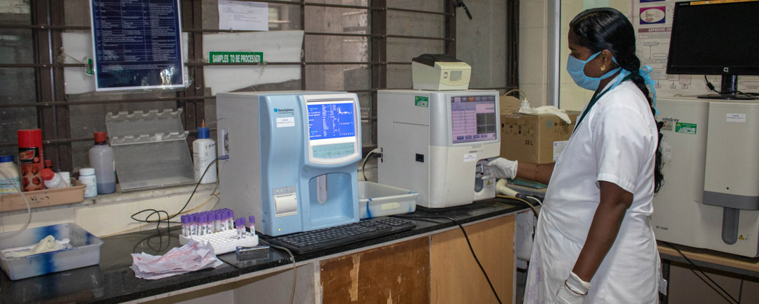
The section of hematology and clinical pathology is located in the SVMCH hospital building as part of the Central laboratory. The section has well trained and capable technicians handling the day to day tests, 24 hours a day, 7 days a week. They are managed by motivated faculty along with postgraduates who are posted to the section in turns. Both internal quality assessments and External Quality Assurance Programs [EQAP] with AIIMS, New Delhiand CMC Vellore are routinely carried out.
Features
- Ø 24/7 hematology and clinical pathology tests
- Ø Dedicated team of faculty & postgraduates
- Ø Technicians on shift duties round the clock
- Ø Adherence to turn around time requirements
- Ø Laboratory Information Software available for reporting the test results
- Ø Emergency/urgent cases handled on first basis
- Ø Internal & external quality checks done periodically
- Ø Equipment are standardized and maintained regularly
Instruments
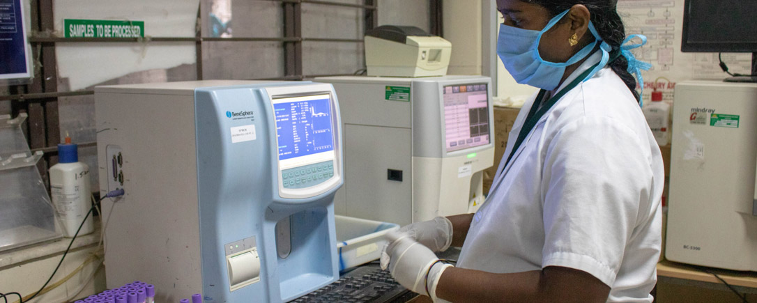
- v BeneSphera H31 (3-part) hematology analyzer-1
- v Mindray BC5150 (5-part) hematology analyzer-1
- v Mindray BC 5300 (5-part) hematology analyzer-1 [fully automated]
- v Coagulation analyzer Ecl105 (Erba)
- v Coagulation analyzer Genius CA51
- v Aimil photochem – calorimeter
- v DiruiH-100 automated urine analyzer
- v Binocular Microscopes –
- v Magnus MLX-DX – 11D452
- v Magnus MLX 11E601
- v Labomed vision 2000
Routine hematology tests
- Complete Blood Count[CBC]/Hemogram Including Haemoglobin, TLC, Differential count, RBC count, Platelet count, AEC, PCV, RDW, MCV, MCH, MCHC
- Peripheral Smear
- MP/MF [Malarial Parasites/Microfilaria]
- ESR [Erythrocyte Sedimentation Rate]
- Reticulocyte count
- Bleeding time/ Clotting time
- Prothrombin time/INR
- APTT [Activated Partial Thromboplastin Time]
- Mixing studies
Specialized tests
- Bone marrow aspiration cytology & biopsy
- Sickling test
- Osmotic fragility test
- Lupus Erythematosus [LE] cell test
Routine clinical pathology tests
- Urine Routine/Complete analysis
- Urine Ketone bodies
- Urine Bile salt& pigment/Urobilinogen
- Urine Bence Jones Protein
- Body Fluids Cell counts
- Semen analysis
- Stool reducing substance
External Quality Assurance/Proficiency testing
- ¤ Hematology – ISHTM-AIIMS EQAP, New Delhi
- ¤ Haemostasis: ISHBT-CMC EQAS, CMC Vellore
SECTION 2: HISTOPATHOLOGY
The histopathology section is situated in the college premises and has the required equipment such as well-ventilated grossing station, semi-automatic tissue processor, rotary microtome, embedding and staining setup and is manned by capable technicians, who are supervised by the departmental faculty. The processed histopathology specimens are reported within an appropriate interval by the trained faculty who in turn train the postgraduates of pathology. When required special tests like additional stains or immunohistochemistry are performed.
Features
- Ø Well-equipped histopathology laboratory
- Ø Routine histopathology processing
- Ø Time bound processing of Small, medium and large surgically resected specimens
- Ø Reasonably accurate & timely Reporting of cases by trained histopathologists
- Ø Communication & coordination with clinicians for satisfactory processing & reporting
- Ø Reports checked by postgraduates and validated by pathologists
- Ø Special stains when required
Instruments
- v Labovision Automatic tissue processor
- v Medimeas Tissue embedding center
- v Leica Semiautomatic microtome
- v Weswox microtome
- v Yorco Cryostat machine
Tests
- Processing of histopathology specimens
- Routine Hematoxylin & Eosin staining
- Special stains/studies available are:
- © Ziehl Neelsen stain
- © Periodic acid Schiff[PAS] stain
- © Giemsa stain
- © Perl’s stain
- © Masson Trichrome stain
- © Van Gieson stain
- © Congo red stain
- © Verhoeff-Van Gieson (VVG) stain
- © Wade Fite stain
- © Methyl violet stain
- © Alcian blue stain
- © Alcian blue – PAS stain
- Facility for Immunohistochemistry
- Frozen section study
SECTION 3: CYTOPATHOLOGY
Cytopathology laboratory is located within the college premisesand managed by an enthusiastic team of full time faculty and postgraduatesin rotation along with qualified and trained technicians, focused on prompt processing and accurate and on-time reporting of cytology samples.
Features
- Ø Fully equipped cytopathology lab
- Ø Set up for FNAC [Fine Needle Aspiration Cytology]
- Ø Processing of FNAC, Fluid samples and Pap smears
- Ø Dedicated faculty, postgraduate and technician
- Ø Timely and accurate reporting of cytopathology cases
Instruments
- v Cytopathology instruments include centrifuge, FNAC gun, staining equipment and routine stains like H&E, Giemsa and Pap stains
Tests
- FNAC – both direct and guided USG/CT guided FNACs are done
- PAP smear –screening test for cervical cancer
- Body fluid cytology – Ascitic, pleural, peritoneal, CSF, synovial fluids are analyzed for inflammation, malignancy etc.


